Document Type : Original Article
Authors
Department of Chemistry, College of Science, Baghdad University, Baghdad, Iraq
Abstract
The titanium dioxide nanoparticles were synthesized by the sol-gel method, while the copper oxide nanoparticles (CuO) were synthesized by the green method by using Sidr extract. The nanocomposites were prepared from the condensation reaction of polystyrene (PS), TiO2, CuO, and (GO) by simple mixing method. These structure (PS/GO/CuO) and (PS/TiO2/CuO) were characterized by FE-SEM, X-rays diffraction, and thermal analysis. The measurements showed that PS/TiO2/CuO and PS/GO/CuO of the synthesized were present in nanoparticle size within the nano-scale. The nanocomposites were tested in the applications of biological activity as antibacterial, antifungal, and antioxidant. The results showed that the nanocomposites (PS/GO/CuO) gave a higher inhibition value than (PS/TiO2/CuO) nanocomposites with bacteria.
Graphical Abstract
)
Keywords
Main Subjects
Introduction
Nanotechnology is one of encouraging development in the current century. The benefit of nanoscience is the ability to transform this science into valuable applications through noticing, control, measurement, and assembly within the nanoscale. In another definition, nanotechnology is a science, technology, and engineering taking place within the nanoscale, it also has applications in all fields of physics, chemistry, medicine, electronics, etc. [1].
Polystyrene is one of the polymers made from its small units of (styrene) (C8H8)n and has a high molecular weight. Polystyrene can be foamed or solid, whilst the monomer styrene is liquid and its non-expansive resin each for unit weight. In addition, polystyrene form a rather poor barrier with water vapor, oxygen, and low melting point [2].
The study of polymer nano composites are creating widely interesting because of their important applications in domestic uses like memory, electronics, and recording head. It causes a change in the physical properties of polymer and implementing novel features for polymer matrix when adding inorganic nanoparticles to the polymers [3].
The oxides of some metallic nanoparticles, such as ZnO, Fe3O4, TiO2, CuO, and CuO are among highly production nanoparticles, so their applications are very wide and utilized as fillers for polystyrene structure, because of their electrical, optical, chemical, and antibacterial properties. As instance, titanium dioxide is low cost and nontoxic material with novel photo catalytic and electrochemical properties generally used the field of chemical fiber production and the UV-resistant material [4].
The NPs green synthesis is an environmentally friendly bio reduction method that does not require the high activation energy needed for the synthesis process. This method has been widely used in the nanomaterial preparation, especially for metal oxides such as actinomycetes and prokaryotic bacteria. Many intracellular proteins and enzymes act as reducing agents for nanoparticle synthesis. Likewise, the other underlying biological agents are plant extracts which are an alternative to conventional processes for preparing nanometal oxides which are inexpensive, simple, and effective. Plant extracts contain biomolecules like proteins, coenzymes, and carbohydrates, which have the ability to reduce mineral salts to nanoparticles [5].
Materials and Methods
Scanning Electron Microscope (SEM) by using “MIRA3 TESCAN: at Taban laboratory/ Iran, Build 20. Transmission Electron Microscope (TEM) by using "FEI Tecnai F20, TF30, JEOL JEM 2100F, FEI Talos F200X: at Taban laboratory/ Iran. X-Ray diffraction (XRD) recorded on ''Philips PW 1730'' portable in PANalytical center/ Iran, 021/44862778. The DSC analyses were measured by using model ''SDT Q600 V20.9 Differential Scanning Calorimetry'' at Taban laboratory/ Iran, Build 20. Thermal gravimetric analysis and differential scanning calorimetry were measured by using model ''SDT Q600 V20.9'' at Taban laboratory/ Iran, Build 20. Magnetic stirrer ''faithful china'' at the University of Baghdad/College of Science/Department of Chemistry Ultrasonic lab disrupter ''BIOBASE UCD-150'' at the University of Al-Mustansiriyah/College of Science. Furnace ''gallenkamp muffle furnace'' at the University of Baghdad/College of Science/Department of Chemistry (Table 1).
Preparation of Sider leaves aqueous extract
A solution of plant extract of Sidr leaves was prepared with a weight of 4 g of plant extract, 150 mL of distilled water was added to it, and then it was heated for 20 minutes to filter the components in the last step [6].
Table 1: The used chemicals materials
|
Ser. |
Name |
Chemical formula |
Purity % |
Company |
|
1 |
Copper(II) sulphate pentahydrate |
CuSO4.5H2O |
99.5 |
CDH |
|
2 |
Titanium isopropoxide |
C12H28O4Ti |
97 |
Sigma-Aldrich |
|
3 |
Sodium nitrate |
NaNO2 |
98 |
CDH |
|
4 |
Potassium permanganate |
KMnO4 |
99.5 |
CDH |
|
5 |
Ammonium Hydroxide |
NH4OH |
99 |
Sigma-Aldrich |
|
6 |
Sulfuric acid (con.) |
H2SO4 |
98 |
Sigma-Aldrich |
|
7 |
Carbon tetra chloride |
CCl4 |
98 |
|
|
8 |
Graphite |
Graphite |
98.2 |
Fasco expoxies |
|
9 |
Hydrogen peroxide |
H2O2 |
97 |
Sigma-aldrich |
|
10 |
Hydrochloric acid (con.) |
HCl |
36.5 |
Sigma-aldrich |
|
11 |
Ziziphus spina-christi Leaf extract |
leaf extracts |
|
Natural |
Synthesis of metal oxides nanoparticles
Copper oxide nanoparticles (Green method) [7]
4 g of Sidr leaf extract was taken; 100 mL of distilled water was added and heated at 80 °C for 15 minutes, after which the solution was filtered. 6 g of copper(II) sulphate pentahydrate (CuSO4.5H2O) was taken and added deionized water to a volume of 250 mL, and then it was heated at 80 °C with stirring for 15 minutes. Sidr leaf extract was added dropwise to CuSO4.5H2O solution with stirring, and then the color was changed to green and deep green. The mixture was heated at 100 °C until all solvent was evaporated. The solution is dried at 60 °C to obtain the precipitate, and then it was calcinated at 400 °C until it became blackish-brown color.
Synthesis of TiO2 nanoparticles [8]
Titanium isopropoxide (10 mL) was dissolved in 100 mL of sider leaves aqueous extract. The mixture was heated at 100 °C for two hours. Yellowish precipitate was found which was then calcined at 350 °C for 2 hours to afford TiO2 NPs.
Synthesis of graphene oxide nanoparticles (Hummers’ method) [9]
1 g of graphite was added very slowly to cool 50 mL of concentrated H2SO4 and it was stirred in an ice bath for 15 minutes. 4 g of sodium nitrate (NaNO2) was added to 6 g of potassium permanganate (KMnO4) to the solution and it was stirred in an ice bath (6 hours). The ice bath was removed and the temperature of the mixture was kept at 35 °C in water path (30 minutes). The mixture became deep red- brown color. 50 mL of deionized water was added to the mixture (in step 4). The temperature was increased to 90- 98 °C, then, the above mixture (in step 5) was diluted by adding 250 mL warm deionized water. 20 mL of H2O2 was added until the mixture turned to bright yellow. The mixture is left for 24 hours, after which the washing process is carried out (to get rid of the acids) until the pH reached 7. The graphite oxide (GO) powder was dried at 40 oC (24 hours).
Synthesis of nanocomposites
Synthesis of (Polystyrene/TiO2/CuO) nanocomposites [10]
25 mL of carbon tetra chloride (CCl4) was added to 1 g of polystyrene and it was refluxed for 1 hour at 50 °C (solution A). 0.1 g of TiO2 NPs and 0.1 g of CuO NPs were mixed in 10 mL of CCl4, and then the mixture was put in ultrasonic for 5 minute (solution B). Solution B was added to solution A, and then it was refluxed for 5 hours. The mixture was collected and left at room temperature until solidified.
Synthesis of (Polystyrene/GO/CuO) nanocomposites [10]
25 mL of CCl4 was added to 1 g of polystyrene, and then it was refluxed for 1 hour at 50 °C (solution A). 0.1 g of GO NPs and 0.1 g of CuO NPs were mixed in 10 mL of CCl4, and then the mixture put in ultrasonic for 10 minutes. (solution B). The solution B was added to the solution A and refluxed for 5 hours. The mixture was collected and left at room temperature until solidified.
Antibacterial activity
Two types of bacteria were selected, they are Gram-negative (Klebsiella pneumoniae) and Gram-positive (Staphylococcus aureus). The bacterial culture process was carried out in a bacterial medium called (Muller Hinton agar). The utilized method is called (well diffusion methods) as follow:
The bacterial culture was prepared by 0.1 mL of 106 CFU/mL broth (liquid medium for culture of bacteria) of indicator strain on the full surface of nutrient agar plate. Three holes were in all the previously prepared agars, the size of each hole was 6 mm. Then, 100 μl of 10 M of the sample (the used solvent was DMSO to prepared all samples) to be studied are injected by micropipette for anti-bacterial activity. Bacterial cultures were incubated at 37 °C for 24 hours, and then the inhibition areas are measured for all compounds [11].
Results and Discussion
X-Ray diffraction of PS/TiO2/CuO nanocomposites
In Figure 1 and Table 2, the X-ray diffraction pattern was demonstrated for PS/TiO2/CuO nanocomposites, the polymer diffraction peaks at 2θ = 19.0702° and 22.683° corresponding to 597.64° and 289.02°, respectively, while it indicated the TiO2 NPs diffraction peaks at 2θ = 25.41°, 26.793°, 48.034°, 54.487°, and 69.729° corresponding to 994.28, 774.58, 341.12, 170.57, and 52.72, respectively, compared with the standard reference for TiO2 NPs [12].
Furthermore, XRD diffraction peaks for CuO NPs at 2θ = were 32.155°, 38.577°, 45.947°, 62.629°, and 75.808° corresponding to 507.61, 209.52, 292.13, 122.24, and 61.98 compared with the standard reference for CuO NPs, as depicted in Figure 2 [13].
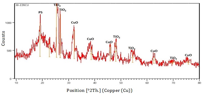
Figure 1: XRD pattern of PS/TiO2/CuO nanocomposites
Table 2: XRD record of PS/TiO2/CuO nanocomposites


Figure 2: XRD pattern of PS/GO/CuO nanocomposites
X-Ray diffraction of PS/GO/CuO nanocomposites
Figure 2 and Table 3 illustrated the X-ray diffraction pattern for PS/TiO2/CuO nanocomposites, polystyrene diffraction peaks at 2θ = 18.5536° corresponding to 331.27°, while XRD diffraction peaks for CuO NPs at 2θ = 35.1124°, 53.9342°, and 72.9231° corresponding to 129.65, 226.23, 226.23, and 233.54, respectively. Furthermore, graphene oxide NPs diffraction peaks at 2θ = 13.8112° and 42.0926° corresponding to 296.32° and 265.01°, respectively [14].
Thermogravimetric analysis of PS/TiO2/CuO nanocomposites
Thermogravimetric analysis of prepared nanocomposites is indicated in Figure 3 in four stages of weight loss [15]:
The first stage: At 35-120 °C with a weight loss percentage of 4.704% was attributed to the loss of water (absorbed from the atmosphere) which physically adsorbed the nanocomposite due to the fact that the nanomaterials have very high surface area.
Table 3: XRD record of PS/GO/CuO nanocomposites


Figure 3: FA electronic spectrum
The second stage: At 120-440 °C and the third stage at 440-800 °C with a total weight (percentage) loss equal to 95.42% (found) corresponding 83.3% (calculated), these stages include the starting of styrene unit loss and there is decomposition of the carbon skeleton of the polymer. The fourth stage: At temperature more than 800 °C with weight (percentage) of 4.566% (found) corresponding 16.6% (calculated), this stage was attributed to the amount of TiO2 and CuO NPs. We noticed that the weight loss at 440 °C of PS/TiO2/CuO nanocomposites was more than that of PS/TiO2/Ag2O nanocomposites (Table 4).
Table 4: All stage decompositions for PS/TiO2/CuO nanocomposites
|
Ser. |
Stage (temperature °C) |
Loss wt% |
Status |
|
1 |
35-120 |
4.704 |
Loss of water |
|
2 |
120-440 |
55.59 |
Starting of loss of styrene unit |
|
3 |
440-800 |
35.14 |
Decomposition of carbon skeleton |
|
4 |
More than 800 |
4.566 |
Metal and metal oxide residue |
Thermogravimetric analysis of PS/GO/CuO nanocomposites
The graphene oxide nanocomposites were thermally decomposed as follows [16]: The first stage: at 35-120 °C with a weight loss percentage of 21.91% was attributed to the loss of water. The second stage: at 120-440 °C and the third stage at 440-800 °C with a total weight (percentage) loss equal to 92.40%, these stages include the starting of styrene unit loss and there is the decomposition of the carbon skeleton of the polymer. The fourth stage: at temperature than 800°C with weight (percentage) of 7.59%, this stage was attributed to the residue amount metal oxides NPs. See Figure 4 and Table 5.

Figure 4: TG analysis of PS/GO/CuO nanocomposites
Table 5: TG analysis of PS/GO/CuO nanocomposites
|
Ser. |
Stage (temperature °C) |
Loss wt% |
Status |
|
1 |
35-120 |
21.91 |
Loss of water |
|
2 |
120-440 |
49.42 |
Starting of loss styrene unit |
|
3 |
440-800 |
21.08 |
Decomposition of carbon skeleton |
|
4 |
More than 800 |
7.59 |
Metal and metal oxide residue |
Differential Scanning Calorimetry (DSC) of (PS/TiO2/CuO) nanocomposites
Differential scanning calorimetry (DSC) analysis was done for the prepared PS/TiO2/CuO and PS/GO/CuO nanocomposites in two endothermic stages: First, at 211.68 °C equal ∆H to 350.9 J/g for PS/TiO2/CuO, at 335.52 °C equal ∆H to 1063 J/g for PS/GO/CuO. It is considered as a glass transition of polystyrene in nanocomposites compared with standard reference of polystyrene which is 100°C. Second, at 252.03 °C and 404.28 °C, respectively that were conceded melting point of polystyrene in nanocomposites compared with the standard reference of polystyrene which is 270 °C [17], as displayed in Figure 5.
The field emission scanning electron microscopy (FE-SEM) of (PS/TiO2/CuO) nanocomposites
The field emission scanning electron microscopy (FE-SEM) measurement indicates that there are two different nano structures, which are irregular sphere like nano structures with diameter range between 45.76-46.27 nm, sheet like nanoparticles with a thickness of approximately 40 nm, these nano structures means that the prepared nanocomposite is within the nanoscale [18] (See Figure 6).


Figure 5: DSC/TGA of a) PS/TiO2/CuO and b) PS/GO/CuO nanocomposites

Figure 6: FE-SEM of PS/TiO2/CuO nanocomposites
The field emission scanning electron microscopy (FE-SEM) of PS/GO/CuO nanocomposites
The field emission scanning electron microscopy (FE-SEM) measurement of the PS/GO/CuO nanocomposites showed that nanoparticles sizes are between 51.56-54.86 nm and this is within the nanoscale. The shape of these nanoparticles is spherical and semi-spherical, as exhibited in Figure 7 [19].
Application
Antibacterial activity
The biological results on bacteria (Klebsiella pneumoniae) and (Staphylococcus aureus) illustrated that PS/TiO2/CuO and PS/GO/CuO nanocomposites have a different effect on inhibiting the growth of the studied bacteria, as presented in Table 6 and Figures 8 and 9. This is due to the ability of these nanocomposites to production of free radicals, which leads to the oxidative stress, and thus damage to proteins, DNA, cell membranes, and binding to cytosolic proteins, DNA, and enzymes. This interaction causes decreased inhibiting respiratory chain, ATP production, and metabolic pathways [20, 21]. The nanocomposites disrupt the act of the bacterial cell membrane through electrostatic binding and release of positively charged metal ions vs. the negative charge of the cell membrane. These charges interfere on the surface through electrostatic binding, which results in an increase in oxidative stress and damage occurs in the cell membrane [22].
Antifungal activity
The anti-fungal activity of PS/TiO2/CuO and PS/GO/CuO nanocomposites synthesized by the green and sol-gel method was studied on Candida Albicans, the results demonstrated that PS/GO/CuO nanocomposites gave the strongest anti-fungal activity against Candida Albicans. This is due to the increase in the surface area of the nanocomposite through the presence of graphene oxide and the small size of copper, which helps to penetrate the cell membrane [23]. (See Table 7 and Figure 10).

Figure 7: FE-SEM of PS/GO/CUO nanocomposites
Table 6: Antibacterial susceptibility test
|
Ser. |
Nanocompsites name |
Staphylococcus aureus (+) |
Klebsiella (-) |
|
1 |
PS/TiO2/CuO |
12 mm |
17 mm |
|
2 |
PS/GO/CuO |
28 mm |
26 mm |

Figure 8: Biological test (Staphylococcus aureus) of PS/GO/Ag2O and PS/TiO2/Ag2O nanocomposites

Figure 9: Biological test (Klebsiella) of PS/GO/Ag2O and PS/TiO2/Ag2O nanocomposites
Table 7: Antibacterial susceptibility test
|
Ser. |
Nanocompsites name |
Candida Albicans |
|
1 |
PS/TiO2/CuO |
16 mm |
|
2 |
PS/GO/CuO |
21 mm |

Figure 10: Biological test of Candida albicans
Corrosion test
The corrosion records were measured and presented in the Table 8 and Figure 11. The measurements indicate that the synthesized nanocomposites gave different results and showed that the carbon steel coated with the synthetic nanocomposites had a high protection rate or inhibition efficiency (IE %) compared with the uncoated carbon steel. The graphene oxides nanocomposites (PS+GO+CuO) gave a high protection rate of 92%, while the titanium dioxide nanocomposites (PS+TiO2+CuO), which amounted to 90%, the recorded also demonstrated an increase in the corrosion resistance value of carbon steel coated with synthetic nanocomposites compared with the used carbon steel [24].
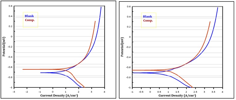
Figure 11: Carbon steel uncoated, PS+TiO2+CuO, and PS/GO/CuO with carbon steel coated
Table 8: Corrosion test
|
Comp. |
E corr. |
I corr. |
I corr./ r |
Resistant |
Anodic β |
Cathodic β |
Corr. rate |
IE% |
|
Blank |
-0.998 |
146.4 |
1.464 |
523.8 |
0.950 |
0.217 |
0.718 |
- |
|
PS+GO+CuO |
-0.666 |
11.90 |
1.190E-5 |
3944 |
0.171 |
0.294 |
0.058 |
92 |
|
PS+TiO2+CuO |
-0.646 |
15.21 |
1.521E-5 |
2816 |
0.140 |
0.332 |
0.075 |
90 |
Conclusion
The nanoparticles synthesis by green methods is an inexpensive (economic) method and at the same time, it preserves the environment from pollution, while the nanoparticles synthesis from chemicals is costly and harmful to the environment. The results proved that the PS+TiO2+CuO and PS+GO+CuO nanocomposites studied by the X-ray diffraction and FE-SEM had the nanoparticles presence of different sizes, within the nanoscale, and the thermal analysis of the nanocomposites was measured and gave an improvement in the physical properties of polystyrene which is an increase in the melting point and glass transition. Biological applications were carried out on the nanocomposites, the tests were conducted on (Staphylococcus aureus) and (Klebsiella) bacteria, the PS+GO+CuOnanocomposite gave the highest inhibition than PS+TiO2+CuO. Furthermore, the antifungal activity was tested on Candida parapsilosis, the test also proved that the PS+GO+CuOnanocomposite is more inhibiting than PS+TiO2+CuO. The results of the anti-corrosion tests gave the protection ratio of PS+GO+CuO nanocomposite (92%) was higher than PS+TiO2+CuO (90%).
Acknowledgements
This research was supported by Dr. Nada Mutter Abbass, university of Baghdad, college of science, department of chemistry. We thank our colleagues who provided insight and expertise that assisted the research. We also do not forget to support the family.
Funding
This research did not receive any specific grant from funding agencies in the public, commercial, or not-for-profit sectors.
Authors' contributions
All authors contributed to data analysis, drafting, and revising of the paper and agreed to be responsible for all the aspects of this work.
Conflict of Interest
There are no conflicts of interest in this study.
ORCID:
Zeyad Zaid Almarbd
https://www.orcid.org/0000-0002-9771-5517
HOW TO CITE THIS ARTICLE
Zeyad Zaid Almarbd, Nada Mutter Abbass. Recycling of plastic waste made of polystyrene and its transformation into nanocomposites by green methods. Chem. Methodol., 2022, 6(12) 940-952
)