Document Type : Review Article
Authors
- Zahra Dourandish 1
- Fariba Garkani Nejad 1
- Reza Zaimbashi 1
- Somayeh Tajik 2
- Mohammad Bagher Askari 3
- Parisa Salarizadeh 4
- Sayed Zia Mohammadi 5
- Hakimeh Oloumi 6
- Farideh Mousazadeh 7
- Mehdi Baghayeri 8
- Hadi Beitollai 1
1 Environment Department, Institute of Science and High Technology and Environmental Sciences, Graduate University of Advanced Technology, Kerman, Iran
2 Research Center of Tropical and Infectious Diseases, Kerman University of Medical Sciences, Kerman, Iran
3 Department of Semiconductor, Institute of Science and High Technology and Environmental Sciences, Graduate University of Advanced Technology, P.O. Box 76318-18356, Kerman, Iran
4 High-Temperature Fuel Cell Research Department, Vali-e-Asr University of Rafsanjan, Rafsanjan 1599637111, Iran
5 Department of Chemistry, Payame Noor University, Tehran, Iran
6 Department of Ecology, Institute of Science and High Technology and Environmental Sciences, Graduate University of Advanced Technology, Kerman, Iran
7 School of Medicine, Bam University of Medical Sciences, Bam, Iran
8 Department of Chemistry, Faculty of Science, Hakim Sabzevari University, Sabzevar, Iran
Abstract
Cancer, is a worldwide epidemic, is characterized by the abnormal growth of cells and their ability to spread to various organs and tissues within the body. Doxorubicin (DOX) is an effective chemotherapy drug that not only inhibits the growth of cancer cells, but also assists in the immune-mediated elimination of tumor cells. Hence, it is critical to carefully regulate the DOX dosage for cancer patients undergoing drug-based cancer treatment. Nowadays, electrochemical sensors have emerged as reliable analytical instruments for detecting a broad spectrum of target molecules. This is because of their simplicity, affordability, and ability to seamlessly integrate with multiplexed and point-of-care strategies. By modifying the surface of electrodes with diverse materials, it is possible to enhance the sensitivity and lower the detection limits (LOD) of electrochemical sensors. This report provides a concise summary of selected studies that focus on the use of electrochemical sensors based on carbon nanomaterials and polymers for the DOX analysis, and offers insights on the technical advancements and potential future applications in this particular domain.
Graphical Abstract
)
Keywords
Main Subjects
Introduction
The continuous advancements in science, technology, and industry have greatly enriched human life. However, as human society progresses and lifespans increase, the prevalence of health issues and diseases poses a significant challenge in modern society [1-3]. Cancer is a prominent disease recognized as the primary cause of global mortality. Different treatment modalities are employed for cancer, and researchers have identified the emergence of a new generation of cancer medications that have undergone advanced clinical trials and demonstrated promising results [4-12].
Doxorubicin, also referred to as hydroxydaunorubicin, is widely recognized as a crucial anti-cancer medication globally [13]. It is classified as a cytotoxic anthracycline drug and is utilized in the treatment of various neoplastic diseases, including acute leukemia, Hodgkin’s and non-Hodgkin’s lymphomas, lung cancer, breast cancer, and sarcomas [14]. DOX functions by interacting with the double helix structure of DNA within cancer cells, specifically targeting the anthracycline moiety. Accordingly, it inhibits the transcription and replication processes of DNA. Unfortunately, the clinical use of DOX is restricted because of the potential development of cumulative dose-dependent chronic cardiomyopathy. This condition, if left untreated, may progress to congestive heart failure, resulting in a mortality rate ranging from 20% to 40% [15,16]. The DOX administration in the human body can lead to various side effects, including systemic toxicity, cardiotoxicity, pain, nausea, and the development of drug resistance during the course of therapy. Therefore, the DOX determination in clinical and biological specimens is very serious due to its significant cardiotoxicity effects [17,18].
To date, several analytical methods such as electrophoresis [19, 20], spectrometry [21], and chromatography [22,23] have been presented for the DOX detection. These strategies are very accurate, but are quite expensive to carry out, time-consuming, laborious, and expensive.
Electrochemical methods are attractive, low cost, fast, portable, no complex pre-treatment, and non-polluting analyses with good kit ability that make them highly attractive compared to the other analytical techniques [24-29]. Electrochemical sensors have become considerable in various fields, including biotechnology and medicine, industrial applications, and environmental monitoring [30-33]. There are 5 types of electro-chemical sensors, i.e. potentiometric, conductive, impedimetric, voltammetric, and amperometric. Among these diverse techniques, amperometric and voltammetric strategies are highly admissible for electro-chemical sensing [34,35].
Electrochemical sensors belong to a class of chemical sensors that utilize an electrode as a transducer to generate an electrochemical signal in response to analytes. Commonly utilized electrodes for electrochemical sensors include glassy carbon, carbon pastes, diamond, gold, graphite, and screen-printed electrodes [36,37]. However, bare electrodes are prone to the adsorption of target analytes and their reducible species during redox reactions. This can lead to contamination of the electrode surface, adversely affecting the analytical reaction performance namely sensitivity, selectivity, and feasibility of redox reactions [38,39].
Modifying the electrodes is considered the most promising approach to enhance various aspects of electrochemical sensing devices, including selectivity, sensitivity, adhesion of analytes, dynamic ranges, and detection limits [40,41]. To enhance sensor performance, various materials are used, including polymer structures, biological elements, as well as conductive and semi-conductive materials [42-45].
Most recently, nanomaterials have been the primary focus of modern research, with a particular emphasis on their potential for modification of electrodes surface [46,47]. Advancements in nanotechnology have enabled the customization of functional nanomaterials through synthetic design, allowing for precise control over their size, composition, and surface properties [48-51]. Nanomaterials possessing remarkable electro-catalytic properties, superior conductivity, and increased surface area have emerged as crucial materials for electrode modification [52-57]. Carbon nanotubes, metal oxide, and metal nano-structures, and graphene oxide are prominent nano-scale materials that have been extensively utilized for electrode modification. They have significantly enhanced the determine process and addressed diverse challenges faced by researchers, such as signal fluctuation and over-potential [58-63].
The principal purpose of this investigation is to develop electro-chemical sensors based on carbon-based nanomaterials and polymer structures.
Electrochemical Sensors Based on Nanomaterials for Doxorubicin Determination
Carbon Nanotubes-Based Electrochemical Sensors for Doxorubicin Determination
Carbon nanotubes (CNTs) were first discovered in 1991 by Sumio Iijima. He discovered CNTs while examining the material that had been deposited on the cathode during the arc-evaporation synthesis of fullerenes [64,65]. Naturally, the CNTs can be categorized into two groups: single-walled carbon nanotubes (SWNTs) and multi-walled carbon nanotubes (MWNTs). SWCNTs exhibit a cylindrical nanostructure, characterized by a high aspect ratio [66, 67].
CNTs are widely recognized as one of the fundamental building blocks of nanotechnology due to their exceptional properties and versatile applications. With a tensile strength approximately 100 times stronger than steel, thermal conductivity better than most materials including diamond, and electrical conductivity comparable to copper but with the capacity to carry higher currents, CNTs are considered an exceptionally captivating material [68-71]. The exceptional properties of CNTs make them incredibly appealing for chemical sensors overall, and electrochemical detection in particular [72,73]. An accurate comparison between the carbon nanotubes-based electrochemical sensors of DOX in terms of analytical figures is presented in Table 1.
Taei et al. fabricated Fe2O3/SnO2 nanocomposite via a simple solid state technique in alkaline medium. Then, they constructed the DNA biosensor for the DOX determination. For this purpose, a combination of MWNTs, Fe2O3/SnO2, and chitosan (CHIT) was immobilized onto a pencil graphite electrode (PGE) surface to enhance the immobilization of double-stranded DNA (ds-DNA) on the electrode surface (ds-DNA- Fe2O3/SnO2-MWNTs-CHIT-PGE). By utilizing the ds-DNA-Fe2O3/SnO2-MWNTs-CHIT-PGE configuration, the researchers could detect the interaction between DOX and ds-DNA. This allowed them to use a DNA-sensor for the susceptible determination of DOX. On the bare PGE surface at pH 7.0, DOX exhibits an oxidation peak at +0.34 V. The DNA presence leads to a decrease in the current, and there is also a positive shift observed in the DOX oxidation peak, which implies an intercalative interaction between DOX and DNA. Finally, the ds-DNA Fe2O3/SnO2-MWNTs-CHIT-PGE sensor exhibits excellent characteristics namely a large detection range (20.0 to 5552.0 nM), good sensitivity, low limit of detection (LOD) (0.004 nM), high stability, rapid response, and good selectivity [74].
In another paper, Taei et al. created a sensor based on an MWCNT/CoFe2O4 nanocomposite-modified carbon paste electrode (MWCNT/CoFe2O4/CPE) and employed it to accurately detect small quantities of DOX using differential pulse voltammetry (DPV). Under optimized experimental conditions, the MWCNT/CoFe2O4/CPE displayed a DPV response at a working voltage of 460 mV which was proportional to the DOX response in the 0.05 to 1150.0 nM range. The LOD was determined to be 10.0 PM. The MWCNT/CoFe2O4/CPE sensor for DOX analysis provides a reliable and efficient method for accurately determining its concentration. This electrode has demonstrated excellent performance and can be applied to the DOX determination in biological specimens [75].
Madrakian et al. used a Fe3O4@Pt nanoparticle and MWCNT modified CPE (Fe3O4@Pt/MWCNT/CPE) as a rapid platform for the voltammetric detection of DOX. The incorporation of MWCNTs and Fe3O4@Pt nanoparticles enhanced the electro-catalytic performance of the developed electrode for determining DOX. The calibration curve was generated using DPV under optimized experimental conditions. It exhibited a linear response range of 0.05 to 70.0 mM for the DOX determination, with a suitable LOD of 1.0 nM. In addition, this method was applied for the voltammetric detection of DOX in urine specimens at low concentrations, yielding satisfactory recovery rates [76].
Haghshenas et al. reported an efficient procedure for creating an electrochemical sensor based on an oxidized MWCNT/glassy carbon electrode (OMWCNT/GCE). The OMWCNT/GCE platform was fabricated using an electrochemical oxidation strategy in a basic medium (0.5 M NaOH solution). It was then utilized as a voltammetric sensor for the simultaneous detection of DOX and dopamine (DA). The sensor displayed remarkable catalytic performance for the oxidation of both DOX and DA. Furthermore, it successfully separated the initially overlapped signals of DOX and DA oxidation on the un-modified electrode, resulting in two distinct and well-defined peaks. Square wave voltammetry (SWV) was engaged to simultaneously detect DOX and DA in a binary mixture. Under optimized conditions, the SWV analysis demonstrated linear concentration dependencies of the anodic current responses for DOX and DA. The concentration range observed was 0.03 to 55.0 μM for DA and 0.04 to 90.0 μM for DOX. The LODs for DA and DOX were determined to be 8.5× 10⁻³ μM and 9.4× 10⁻³ μM, respectively. The practical applicability of the OMWCNT/GCE was further demonstrated via successfully detecting DOX and DA simultaneously in urine and blood serum specimens [77].
Hajian et al. developed an electrochemical platform utilizing a platinum electrode modified with MWCNTs (Pt/MWCNTs) for the DOX determination, a chemotherapy drug, in plasma samples. DOX was successfully adsorbed on the Pt/MWCNTs surface, resulting in the appearance of a pair of redox peaks at approximately 0.522 V and 0.647 V in 0.1 M Britton Robinson buffer (B-R) at a pH of 4.0. The electrochemical parameters, containing pH, accumulation time, buffer type, and amount of modifier were optimized in this study. Under the ideal conditions, a linear calibration curve was observed within the 0.05 to 4.0 μg/mL range. The LOD for DOX was achieved to be 0.002 μg/mL [78].
Kalambate et al. introduced a voltammetric platform for the simultaneous detection of the DOX and dasatinib (DAS) (as anticancer drugs). This sensor utilized a GCE modified with mesoporous Pd@Pt core-shell supported on MWCNT (Pd@Pt/MWCNT/GCE). The electrochemical behavior of the DAS and DOX was investigated using cyclic voltammetry (CV), while simultaneous detection was performed via adsorptive stripping square wave voltammetry (AdSSWV). The developed sensor exhibited exceptional electrochemical response to DAS and DOX within the linear concentration ranges of 38.0-9880.0 nM and 4.4-8580.0 nM, respectively. The LOD for DOX was computed to be 0.86 nM, while for DAS it was determined to be 6.72 nM. Moreover, the developed sensor was engaged for the accurate and precise detection of DAS and DOX in urine and blood serum specimens. This demonstrates the reliability and practicality of the platform for real-world applications in biomedical analysis [79].
Sharifi and Fayazfar investigated a GCE modified with MWCNTs decorated with Au nanoparticles (MWCNTs/AuNPs/GCE) as an ultrasensitive sensing platform for the detection of DOX.
Figure 1 illustrates the fabrication strategy of the MWCNTs/AuNPs/GCE. The strategic combination of MWCNTs and Au NPs resulted in a synergistic effect that enhanced the rate of electron transfer. This synergistic effect enabled the development of an active platform for sensitive detection of DOX. Also, a broad concentrations range of DOX from 1×10-11 to 1×10⁻⁶ M was achieved using linear sweep voltammetry (LSV) at the modified electrode. In addition, a very low LOD of 6.5 pM was obtained, demonstrating the excellent sensitivity of the method. The fabricated sensor exhibited enhanced electro-catalytic activity, repeatability, and high stability. It also demonstrated satisfactory selectivity for detecting DOX [80].
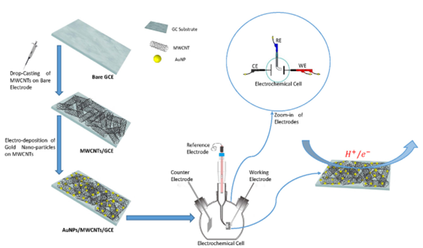
Figure 1: Schematic representation of the MWCNTs/AuNPs/GCE sensor [80]
Zhao et al. introduced a novel electrochemical platform for the DOX detection. The sensor utilized a covalent organic framework decorated with AuNPs and MWCNTs (AuNPs@COFs-MWCNTs) as the modifier material. The AuNPs@COFs-MWCNTs nanocomposite was utilized to modify the GCE surface (AuNPs@COFs-MWCNTs/GCE) through a simple physical deposition method. This modification process resulted in a significantly amplified response signal for DOX detection.
The porous nature and high surface area of COFs enabled better distribution of electro-active sites and enhanced affinity towards DOX in the AuNPs@COFs nanocomposite. This, in turn, enhanced the electrocatalytic activity of the nanocomposite towards DOX detection. Furthermore, highly conductive MWCNTs were incorporated into the AuNPs@COFs nanocomposite to ensure optimal conductivity. As a result, the AuNPs@COFsMWCNTs nanocomposite led to improved catalytic activity which extremely amplified the response signal for DOX detection. Consequently, the developed electrode demonstrated an extended linear range for DOX detection, spanning from 0.08 μM to 25.0 μM. Moreover, it achieved a low LOD of 16.0 nM, indicating the high sensitivity of the sensor for DOX detection. It effectively detected DOX in spiked cell lysate and human serum samples, indicating its practical application for monitoring DOX drug levels in a clinical setting [81].
Table 1: Comparison of analytical figure for electrochemical detection of DOX using carbon nanotubes-based electrochemical sensors
|
Electrochemical Sensor |
Electrochemical Method |
Limit of Detection |
Linear Range |
Ref. |
|
ds-DNA- Fe2O3/SnO2-MWCNTs-CHIT-PGE |
DPV |
0.004 nM |
20.0 to 5552.0 nM |
[74] |
|
MWCNT/CoFe2O4/CPE |
DPV |
10.0 pM |
0.05 to 1150.0 nM |
[75] |
|
Fe3O4@Pt/MWCNT/CPE |
DPV |
1.0 nM |
0.05 to 70.0 mM |
[76] |
|
OMWCNT/GCE |
SWV |
9.4 nM |
0.04 to 90.0 μM |
[77] |
|
Pt/MWCNTs |
CV |
0.002 μg/mL |
0.05 to 4.0 μg/mL |
[78] |
|
Pd@Pt/MWCNT/GCE |
AdSSWV |
0.86 nM |
4.4–8580.0 nM |
[79] |
|
MWCNTs/AuNPs/GCE |
LSV |
6.5 pM |
0.01 to 1000.0 nM |
[80] |
|
AuNPs@COFs-MWCNTs/GCE |
DPV |
16.0 nM |
0.08 to 25.0 μM |
[81] |
Graphene-Based Electrochemical Sensors for Doxorubicin Determination
Graphene, a two-dimensional (2D) lattice comprised of single-atom-thick nano-structured sheets arranged in a honeycomb pattern, is a prominent member of the carbon nanoscale materials family [82,83]. In 2004, Geim and Novoselov conducted an experimental study on the exfoliation, electronic properties, and characterization of this 2D carbon via repeatedly cleaving graphite using adhesive tape [84]. Due to its intrinsic and unique mechanical and electronic properties, graphene is further utilized as a material in a broad spectrum of applications [85-90]. The significant advantages of graphene-based materials such as mass production, high surface area, superior conductivity, low cost, chemical and thermal stabilities properties, and wide potential window have promoted their further applications for electrochemical catalysis and sensing [91,92]. An accurate comparison between the graphene-based electrochemical sensors of DOX in terms of analytical figures is summarized in Table 2.
Guo et al. suggested an ultra-sensitive sensor for the determine DOX and methotrexate using a GCE modified with the cyclodextrin-graphene nanosheets (CD-GNs/GCE). The electrochemical response of DOX and methotrexate at the proposed electrode demonstrated significantly improved electrochemical responses compared to that at the un-modified GCE. The hybrid nanomaterial greatly enhanced the electrochemical response of both drugs by harnessing the respective advantages of cyclodextrin and graphene in the sensor design. The electrochemical sensor demonstrated linear response ranges of 10.0 nM-0.2 mM for DOX and 0.1-1.0 mM for methotrexate. The LODs for DOX and methotrexate were determined to be 0.1 nM and 20.0 nM, respectively. The properties exhibited by the fabricated sensor make it a good platform for the accurate determination of DOX and methotrexate in various domains such as clinical, biology, and pharmaceutical fields [93].
Chekin et al. discussed the design and development of a disposable electrochemical sensor that can be used for directly monitoring DOX levels in clinical blood specimens. The researchers utilized a gold electrode coated with the nitrogen-doped reduced graphene oxide (N-rGO) and chitosan, resulting in a sensor denoted as Au/N-rGO-CS. By optimizing the experimental conditions, the researchers established a linear correlation between the anodic current and the DOX level within the range of 0.010-15.0 μM. The sensor demonstrated an LOD of 10.0 nM, indicating its sensitivity to low concentrations of DOX. The offered sensor was engaged for the DOX determination in serum specimens obtained from patients undergoing anti-cancer treatment [94].
Lee et al. demonstrated the production of high-quality graphene nano-sheets through liquid-phase shear exfoliation in organic solvents, specifically 1-methyl-2-pyrrolidinone (NMP), at ambient conditions, and then urea was introduced as a stabilizer for this process. They obtained the low-defect graphene (LDG) utilizing this method, which is rather straightforward and accessible, thereby rendering it an efficient way for large-scale production of graphene. In addition, the researchers used the LDG to modify a GCE (LDG-GCE) to develop an electrochemical sensor for DOX. The proposed sensor exhibited improved electro-catalytic activity towards DOX, resulting in a high sensitivity of 7.23 × 10⁻¹ μM/μA. It also achieved a low LOD of 39.3 nM [95].
Yan et al. presented a simple and efficient approach for integrating a vertically-ordered mesoporous silica-nanochannel film (VMSF) with electrochemically reduced graphene oxide (ErGO). This integration was achieved using an electrochemically assisted self-assembly approach. Electrochemical reduction of GO and growth of the VMSF both take place simultaneously in a straightforward one-step process, forming a VMSF/ErGO layer on the GCE (VMSF/ErGO/GCE). Due to the presence of oxygen groups, 2D planar structure, and the hydrophobic structure (π-conjugated) of ErGO, the VMSF was able to grow on the GCE surface stably. This layer further served as a protective barrier, preventing the internal ErGO electro-active layer from detaching from the surface of electrode after prolonged usage. Compared to an un-modified GCE, the VMSF/ErGO/GCE demonstrated superior characteristics in detecting DOX. It displayed a linear range of 1.0 nM to 20.0 mM, a good sensitivity of 7.815 mA mM-1, and a good LOD of 0.77 nM. These exceptional results were achieved through the combined signal amplification effects of the electro-catalytic activity and π–π interaction provided by ErGO, as well as the electrostatic preconcentration effect offered by the VMSF [96].
Shi et al. successfully synthesized 3D nanoflower-like ZnO-graphene oxidation nanocomposites (3D ZnO-GO) using a straightforward aqueous hydrothermal approach and a sonochemical method. Afterwards, the researchers proceeded to decorate Au@AuPt nanoparticles onto the 3D ZnO-GO, resulting in the creation of novel Au@AuPt/3D ZnO-GO nanohybrids. These nanohybrids were then utilized to construct an electrochemical sensor for the determination of DOX (Au@AuPt/3D ZnO-GO/GCE). Compared to un-modified electrode, the Au@AuPt/3D ZnO-GO/GCE demonstrated a notable improvement in the current response. The created sensor indicated a broad linear concentration range of detection, spanning from concentrations as low as 0.65 μM up to 369.45 μM, with a low LOD of 0.013 μM. In addition, the developed electrode was utilized for the DOX determination in real specimens (urine) [97].
Rezvani Jalal et al. developed a voltammetric sensor by utilizing the in situ growth of NiCo-BTC bimetallic Metal-Organic Frameworks (MOFs). These MOFs were grown on a GCE, which had been previously modified with conductive nitrogen-doped GO nanoribbons (NiCoBTC MOFs/N-GONRs/GCE). The SWV response of the NiCo-BTC MOFs/N-GONRs/GCE toward DOX showed a significantly higher signal compared to NiCoBTC MOFs/GCE. This improvement can be attributed to the synergistic effect from NiCo-BTC MOFs and N-GONRs. Under optimal conditions, the developed electrode exhibited a powerful current response to the DOX oxidation. The calibration curve generated for DOX using the proposed sensor exhibited two linear ranges: 0.01-1.0 μM and 1.0-80.0 μM. The LOD was determined to be 0.006 μM (or 6.0 nM). This detection limit is lower than the DOX concentration typically found in human plasma specimens, which is approximately 77.2 ± 10.5 nM. The results obtained indicate that the developed sensor holds great promise for accurately determining DOX concentrations in serum and human urine specimens [98].
Table 2: Comparison of analytical figure for electrochemical detection of DOX using graphene-based electrochemical sensors
|
Electrochemical Sensor |
Electrochemical Method |
Limit of Detection |
Linear Range |
Ref. |
|
CD-GNs/GCE |
DPV |
0.1 nM |
10.0 Nm to 0.2 mM |
[93] |
|
Au/N-prGO-CS |
DPV |
10.0 nM |
0.010 to 15.0 μM |
[94] |
|
LDG‐GCE |
DPV |
39.3 nM |
0.3 to 3.0 μM |
[95] |
|
VMSF/ErGO/GCE |
DPV |
0.77 nM |
1.0 nM to 20.0 mM |
[96] |
|
Au@AuPt/3D ZnO-GO/GCE |
DPV |
0.013 μM |
0.65 to 369.45 μM |
[97] |
|
NiCoBTC MOFs/N-GONRs/GCE |
SWV |
0.006 μM |
0.01 to 80.0 μM |
[98] |
Other Carbon Nanomaterials-Based Electrochemical Sensors for Doxorubicin Determination
An accurate comparison between the other carbon nanomaterials-based electrochemical sensors of DOX in terms of analytical figures is listed in Table 3.
Hasanzadeh et al. prepared a GCE modified with graphene quantum dots (GQDs) using casting GQDs suspension onto its surface (GQD-GCE). This electrode was then employed for the detection of DOX in plasma specimens. It was discovered that GQD had been stably absorbed on the GCE using a straightforward procedure. The results obtained from CV experiments revealed a significant enhancement in electro-activity for the DOX oxidation in phosphate buffer solutions (PBS) when using the GQD-modified GCE. The linear range of concentration for the detection of DOX using the GQD-modified GCE was found to be 0.018-3.60 μM. The LOD achieved under these optimized conditions was determined to be 0.016 μM [99].
Table 3: Comparison of analytical figure for electrochemical detection of DOX using other carbon nanomaterials -based electrochemical sensors
|
Electrochemical Sensor |
Electrochemical Method |
Limit of Detection |
Linear Range |
Ref. |
|
GQD-GCE |
DPV |
0.016 μM |
0.018 to 3.60 μM |
[99] |
|
CuNPs-CB-Nafion/GCE |
SWV |
0.024 μM |
0.45 to 5.1 μM |
[100] |
|
GCE/N-CNOs |
DPV |
60.0 pM |
0.2 nM to 10.0 µM |
[101] |
|
FeV/SCNFs/GCE |
Amperometric |
5.2 nM |
0.02 to 542.5 µM |
[102] |
|
CDs/CeO2/SPCE |
CV |
0.09 μM |
0.2 to 20.0 μM |
[103] |
Materon et al. proposed a voltammetric platform for the simultaneous detection of DOX and methotrexate by Cu nanoparticles (CuNPs), Nafion, and carbon black (CB), modified GCE (CuNPs-CB-Nafion/GCE). The combination of CB and CuNPs in the CuNPs-CB-Nafion/GCE sensor resulted in outstanding catalytic performance for the detection of both DOX and methotrexate. The proposed sensor demonstrated excellent catalytic activity for electrochemical oxidation, as evidenced by the SWV results. The redox potentials observed for DOX and methotrexate were 0.69 V and 0.93 V, respectively, indicating efficient oxidation of both analytes. Upon optimization, the CuNPs-CB-Nafion/GCE sensor demonstrated a linear range of 4.5 × 10⁻⁷ to 5.1 × 10⁻⁶ M for DOX, with a LOD of 2.4 × 10⁻⁸ M. Similarly, for methotrexate, the linear concentration range achieved was 2.2 × 10⁻⁶ to 2.5 × 10⁻⁵ M, with a LOD of 9.0 × 10⁻⁸ M. The created sensor proved to be effective in the determination of DOX and methotrexate in biological matrices, such as urine specimens, as well as environmental samples, like water river samples. The sensor exhibited a spike recovery rate of nearly 100%, indicating its accuracy and reliability in these real sample matrices [100].
Ghanbari and Norouzi developed a sensor for the DOX determination using a GCE that was modified with nitrogen-doped carbon nanoonions (GCE/N-CNOs). They prepared the N-CNOs from the fullerene via a straightforward procedure utilizing aminated nanodiamonds (AM-NDs). During the preparation process, nitrogen atoms were introduced into the CNO cages via annealing the AM-NDs under an inert atmosphere and reduced pressure. This allowed for the incorporation of nitrogen into the nanostructures. The results of the study revealed that the N-CNOs possessed intriguing physicochemical properties. These N-CNOs exhibited a high active surface area, measuring 1.41 cm2, as well as excellent electro-catalytic activity. These properties made the N-CNOs an ideal choice for sensor construction, as they provided an active site for the DOX determination. The GCE/N-CNOs displayed a linear response for the DOX detection in a concentration range of 0.2 nM to 10.0 µM. The sensor achieved a low LOD of 60.0 pM, and a calculated sensitivity of 1.13 µA µM-1 cm-2. To assess the practicality of the DOX sensor, a blood serum sample was applied for testing [101].
Rajaj et al. developed a composite material consisting of iron vanadate nanoparticles assembled with sulfur-doped carbon nanofibers (FeV/SCNF). The FeV/SCNFs electrocatalyst was then modified onto a GCE to create FeV/SCNFs/GCE, which was employed for the DOX detection. The FeV/SCNFs/GCE demonstrated the excellent sensitivity (46.041 μA μM-1 cm-2) within a broad concentration range of 20.0 nM to 542.5 μM. Likewise, the sensor indicated superior selectivity even in the presence of common interferents, making it suitable for accurate and reliable DOX detection. Moreover, the FeV/SCNFs/GCE was engaged to determine DOX in diverse real specimens. Specifically, the determination of DOX in human urine and blood serum was performed, and the accepted results demonstrated a recovery range of 98.38% to 99.92%. This indicates the reliability and accuracy of the FeV/SCNFs-based sensor in real specimens’ analysis [102].
Thakur et al. introduced a novel modification to a screen-printed carbon electrode (SPCE) by incorporating carbon dots/ CeO2 nanocomposites (CDs/CeO2). The resulting modified electrode, termed CDs/CeO2/SPCE, was designed for the sensitive determination of DOX. Therefore, they initially synthesized CeO2NPs from urea and (NH4)2[Ce(NO3)6] using easy refluxing, and then CDs were fabricated utilizing taurine using the thermal decomposition technique. After that, CDs/CeO2 were prepared with various wt% of CDs (from 0.5 to 5 wt%) through a hydrothermal approach. The CDs/CeO2 displayed more efficiency for DOX sensing compared to bare CDs and CeO2NPs via facilitating electron transfer response at the surface of SPCE with increasing amounts of CDs. The fabrication process of the nanocomposites and modified electrode for detection of DOX is depicted in Figure 2. The 5wt% CDs-5.0/CeO2 nanocomposite exhibited the most elevated oxidation reaction towards 20 μM of DOX (pH=5.0). The CV revealed that the CDs-5.0/CeO2/SPCE showed a linear response range of 0.2-20.0 μM, and a low LOD of 0.09 μM for DOX oxidation [103].
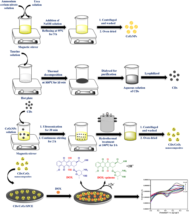
Figure 2: A schematic diagram for the synthesis of CeO2NPs, CDs, and CDs/CeO2 nanocomposites, and electrochemical detection of DOX [103]
Polymers-Based Electrochemical Sensors for Doxorubicin Determination
Nowadays, a broad spectrum of compounds being designed is polymers. Concerning the diversity of their physical and chemical properties, they can adapt to many applications [104-114]. Most recently, enormous attraction has been established in polymeric materials that could irreversibly or reversibly change their chemical and physical properties under the influence of foreign stimuli, including temperature, pH, light radiation, presence of specific ions, magnetic fields, mechanical forces, bioactive molecules, and electric fields [115-124].
The study of polymer films on the electrodes surface is currently one of the most dynamic areas of research in the field of modern electrochemistry. The modification of polymeric species through adsorption or coating onto electrode surfaces offers significant flexibility. Polymers with various functional groups can achieve substantial surface coverage through thick multilayer coatings [125]. This characteristic facilitates the attachment of certain compounds to the polymer matrix-coated electrodes, allowing them to mediate the oxidation of electro-active species. Among the various techniques for creating polymeric-modified electrode, electro-polymerization has emerged as an efficient and versatile method due to its benefits such as strong adherence to the electrode surface and good chemical stability of the film analysis, superior selectivity, sensitivity and reduced costs, and homogeneity in electrochemical deposition [126-128]. An accurate comparison between the polymers-based electrochemical sensors of DOX in terms of analytical figures is presented in Table 4.
Table 4: Comparison of analytical figure for electrochemical detection of DOX using polymers -based electrochemical sensors
|
Electrochemical Sensor |
Electrochemical Method |
Limit of Detection |
Linear Range |
Ref. |
|
PGA-GCE |
SWV |
0.45 µM |
2.20 to 44.5 µM |
[129] |
|
GCE/Poly(Neutral red)/ thiacax[4] arene/DNA |
DPV |
0.05 nM |
0.1 to 100.0 nM |
[130] |
|
Impedimetric |
0.1 nM |
0.01 to 100.0 µM |
||
|
PARG-GCE |
DPV |
69.0 nM (whole blood) 103.0 nM (plasma) |
0.069 to 1.08 µM (whole blood) 0.1 to 3.45 μM (plasma) |
[131] |
|
PANI/DNA/PANI/GCE |
Impedimetric |
0.6 pM |
1.0 pM to 0.1 µM |
[132] |
|
GCE/Poly(Azure B–proflavine)/DNA |
Impedimetric |
0.01 nM |
0.03 to 10.0 nM |
[133] |
|
GCE/poly-proflavine /DNA |
Impedimetric |
0.3 nM |
1.0 nM to 0.1 μM |
[134] |
|
GCE/poly-Azure B |
Impedimetric |
0.07 nM |
0.1 µM to 0.1 nM |
[135] |
|
PEGylated-CoFe2O4/GCE |
DPV |
- |
30 ng/mL to 5.0 μg/mL |
[136] |
Santos et al. used the Poly-L-glutamic acid (PGA), a biodegradable polymer, as conjugated to DOX and also in the modification GCE (PGA-GCE). The interaction occurs between the carboxyl groups of the PGA film and amino groups of the DOX drug, and it serves as the foundation for the development of a straightforward sensor for DOX detection. DOX pre-concentration takes place on the PGA-GCE under open circuit conditions and is analyzed using the SWV method to track the target drug. The calibration curves generated were linear within the range of 2.20 to 44.5 µM, and a LOD of 0.45 µM was achieved. These outcomes demonstrate that the fabricated electrode is appropriate for determining DOX in real specimens [129].
Evtugyn et al. offered the development of a DNA sensor for detecting anthracycline preparations. The sensor was based on a GCE modified with polycarboxylated thiacax[4]arene and electropolymerized Neutral red (NR), which had a mediator covalently attached and DNA electrostatically adsorbed onto it. This sensor, referred to as GCE/Poly(neutral red)/thiacax[4]arene/DNA, showed high sensitivity in detecting anthracycline preparations. The intercalation of DOX, idarubicin, and daunorubicin, into DNA causes an increase in charge transfer resistance and a decrease in electron exchange. This leads to decay in the cathodic peak of NR reduction. As a result of these changes, it became possible to accurately determine concentrations as low as 0.1 nM for daunorubicin, 0.05 nM for DOX, and 0.5 nM for idarubicin. In addition, the DNA sensor was tested for detecting DOX in artificial blood plasma and pharmaceuticals, yielding recovery rates of 95-100% [130].
Soleymani et al. applied a poly-arginine thin film on a GCE (PARG-GCE) via a step electrodeposition process to determine DOX in clinical samples. The CV results revealed that the DOX oxidation occurs via the participation of two electrons and protons at a pH=7.0, as detected by the PARG-GCE sensor.
Furthermore, a significant aspect of the study is the occurrence of electrostatic repulsion between the PARG-GCE and the specific drug, leading to the signal amplification upon the DOX oxidation. This process also reduces the over-potential of DOX, enabling the determination of DOX in real specimens. Moreover, by employing the DPV approach, the DOX detection in plasma and whole blood specimens was gained. The lower limit of quantification for DOX in whole blood was calculated to be 69.0 nM, while in plasma samples it was determined to be 103.0 nM. The findings demonstrated that this sensor has the potential to be utilized for real-time and online monitoring of DOX, an important anticancer drug, in real specimens [131].
Kulikova et al. introduced a DNA sensor utilizing a platform consisting of a GCE modified with DNA sandwiched between two electro-polymerized layers of polyaniline (PANI/DNA/PANI/GCE). The surface layer was constructed through sequential steps involving potentiodynamic electrolysis, DNA drop casting, and a second round of electrolysis. This second electrolysis step was crucial, as it encapsulated the DNA molecules, preventing their leaching into the solution. To measure DOX, the DNA-sensor was initially incubated in a solution of Methylene blue. This step amplified the signal by facilitating DNA intercalation and creating competition between Methylene blue and DOX for the available DNA binding sites. The calibration curve developed was linear within the range of 1.0 pM to 0.1 µM. The DNA sensor was tested to monitor artificial urine specimens, demonstrating acceptable recovery rates [132].
Porfireva and Evtugyn introduced a DNA sensor for DOX detection utilizing a GCE modified with electro-polymerized Azure B and proflavine, along with the adsorption of native DNA from salmon sperm onto a polymer film (GCE/Poly(Azure B-proflavine)/DNA). The investigations revealed a distinction in the behaviour between the individual drugs when polymerized and when in a mixture. The value of the charge transfer resistance exhibited a consistent increase corresponding to the DOX concentration within the range of 0.03 to 10.0 nM, with a LOD of 0.01 nM. The DNA sensor was subjected to testing using DOX preparations and spiked specimens mimicking blood serum. The recovery rate was determined to be 98-106%, indicating the accurate and reliable performance of the DNA sensor in detecting DOX in these samples [133].
Porfireva et al. utilized the electro-polymerization of proflavine to physically adsorb native DNA, followed by the measurement of anthracycline drugs (daunorubicin and DOX) capable of intercalating with DNA. The redox properties of the proflavine polymers on the GCE and DNA deposition on the GCE/poly-proflavine platform (GCE/poly-proflavine /DNA) were described utilizing approaches such as CV, and scanning electron microscopy. As a result, when the GCE/poly-proflavine /DNA sensor was incubated in the drug solution, it led to an increase in the charge transfer resistance. The impedimetric response demonstrated a consistent increase corresponding to the concentration of drugs within the range of 1.0 nM to 0.1 μM for DOX and 1.0 pM to 10.0 nM for daunorubicin. The LOD for DOX was calculated to be 0.3 nM, while for daunorubicin it was determined to be 0.001 nM [134].
Porfireva et al. developed a voltammetric DNA sensor for the determine DOX utilizing a GCE modified with an electropolymerized film of Azure B, along with the physical adsorption of native DNA (GCE/poly-Azure B). The redox behaviour of the polymeric Azure B was investigated at different pH levels and scan rates to monitor its behaviour. Under optimal conditions, the DNA sensor enables the detection of DOX within the range of 0.1 µM to 0.1 nM, with a LOD of 7 × 10-11 M. This sensor was subjected to testing using commercial DOX formulations as well as artificial specimens mimicked the electrolyte content of human serum. A recovery rate of approximately 90% was observed, indicating the reliable performance of the DNA sensor in accurately detecting DOX in these samples [135].
Abbasi et al. fabricated a sensor by employing tryptophan (Trp) and polyethylene glycol (PEG)-functionalized CoFe2O4 NPs to modify the GCE surface. The fabricated electrode (PEGylated-CoFe2O4/GCE) was applied for the determine DOX in unprocessed plasma specimens. The incorporation of PEG molecules into the electrode design provided an antifouling effect, which served to inhibit the precipitation of macromolecules on the prepared electrode surface. Figure 3 demonstrates the fabrication steps of DOX sensor. The designed sensor exhibited exceptional catalytic activity for the DOX oxidation due to the increased conductivity and the presence of electro-catalytic active sites. This enhanced catalytic effect facilitated the efficient and accurate detection of DOX. After optimizing the sensor conditions, the proposed sensor achieved a low limit of quantification of 30 ng/mL for the DOX determination. In addition, the linear range for the detection of DOX was found to be 30 ng/mL to 5.0 μg/mL [136].
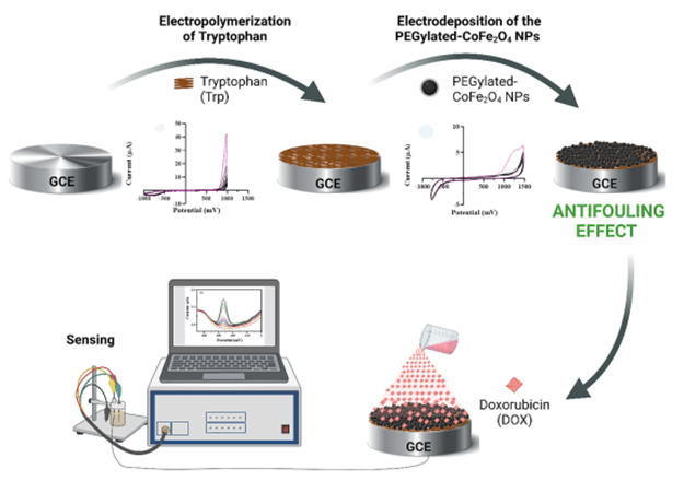
Figure 3: Scheme of DOX sensor designing steps [136]
Conclusion
Doxorubicin is a chemotherapy medication used for the treatment of diverse kinds of cancer. Recent surveys have shown that cardiomyopathies and myelosuppression are associated with the utilization of high doses of DOX. Therefore, the DOX determination in clinical and biological specimens is influential due to its significant cardiotoxic effects. Among different instrumental techniques, electrochemical sensor systems have gained more popularity. This can be attributed to their field-portable capabilities and simpler instrumentation requirements, ultimately leading to reduced costs. A variety of electrochemical sensors based on carbon nanostructures and polymer structures used for detection of DOX are presented in this review.
Choosing a suitable electrode material is a key challenge in the development of electrochemical sensors. Understanding the molecular-level connection between surface structure and reactivity is crucial for sensor design. Having knowledge about interfacial reaction kinetics and sensing mechanisms plays an essential role in designing sensors with improved sensitivity, selectivity, and lower detection limits.
Based on the findings, it can be concluded that modification of the electrode surface using graphene and CNTs has led to an enhancement in surface area and porosity. Furthermore, carbon-based materials such as graphene and CNTs demonstrate effective quantification capabilities and are particularly appealing due to their lower cost compared to noble metals. In addition, it has been discussed that the polymer material contributes to providing conductivity, while the complex ligand used for functionality serves as a strategic component in the overall system.
Hence, future research on this electrode material should concentrate on gaining a deeper understanding of interfacial reaction kinetics to develop innovative sensors that are appropriate for a wide range of practical applications. Moreover, as the demand for point-of-care testing grows, there is a greater emphasis on miniaturized devices. Such miniaturized analytical instruments not only decrease the volume of liquid being processed, but also offer quicker analysis and lower operational costs.
ORCID
Hadi Beitollahi
https://orcid.org/0000-0002-0669-5216
HOW TO CITE THIS ARTICLE
Dourandish, F. Garkani Nejad, R. Zaimbashi, S. Tajik, M. Bagher Askari, P. Salarizadeh, S. Z. Mohammadi, H. Oloumi, F. Mousazadeh, M. Baghayeri, H. Beitollahi. Recent Advances in Electrochemical Sensing of Anticancer Drug Doxorubicin: A Mini-Review. Chem. Methodol., 2024, 8(4) 293-315
OPEN ACCESS
©2024 The author(s). This article is licensed under a Creative Commons Attribution 4.0 International License, which permits use, sharing, adaptation, distribution and reproduction in any medium or format, as long as you give appropriate credit to the original author(s) and the source, provide a link to the Creative Commons license, and indicate if changes were made. The images or other third party material in this article are included in the article’s Creative Commons license, unless indicated otherwise in a credit line to the material. If material is not included in the article’s Creative Commons license and your intended use is not permitted by statutory regulation or exceeds the permitted use, you will need to obtain permission directly from the copyright holder. To view a copy of this license, visit: http://creativecommons.org/licenses/by/4.0/
PUBLISHER NOTE
Sami Publishing Company remains neutral concerning jurisdictional claims in published maps and institutional affiliations.
CURRENT PUBLISHER
)